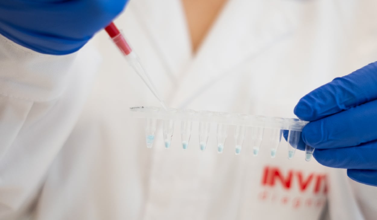
Breast Cancer Awareness Month offers an opportunity to examine the laboratory workflows supporting cancer research and molecular diagnostics.
Breast cancer illustrates the complexity of modern molecular oncology particularly clearly. Depending on the question, laboratories may investigate inherited cancer-predisposition variants, tumour-specific DNA alterations, gene-expression patterns, or molecular mechanisms associated with treatment response and resistance.
The same principles apply across oncology. Whether laboratories are investigating inherited cancer risk, profiling tumour DNA, studying RNA expression, examining virus-associated cancers or developing assays for specific biomarkers, the quality of the result is closely linked to what happens before PCR, sequencing or other analytical methods begin.
Appropriate sample collection, stabilisation and nucleic acid extraction influence whether material entering an assay is representative, sufficiently intact and suitable for the intended analysis.
In this InviBlog article, we explore a range of molecular oncology applications, from hereditary cancer testing and tumour profiling to RNA analysis, virus detection and biomarker-assay development, and examine where Invitek products and services can support key workflow steps.
1. Hereditary cancer testing: understanding germline variation
Some people inherit genetic variants that increase their risk of developing particular cancers. Variants in BRCA1 and BRCA2 are among the best-known examples, with relevance to breast, ovarian, prostate and pancreatic cancer. Other hereditary cancer syndromes involve genes linked to colorectal, endocrine and several additional cancer types [1].
These investigations focus on germline DNA: DNA inherited from parents and present in most cells of the body. Whole blood and buffy coat are commonly used sources. Saliva and buccal swabs can provide practical, non-invasive alternatives, particularly when collection takes place outside the laboratory or samples are gathered from larger or geographically dispersed groups.
The type of genetic change being investigated matters. Some workflows examine a known single-base change, commonly described as a single-nucleotide variant, or SNV. When such a variant is relatively common in a population, it may be referred to as a single-nucleotide polymorphism, or SNP. Many SNPs have no known clinical effect, while some variants may be associated with cancer susceptibility, pharmacogenetic response or ancestry [2].
In hereditary cancer testing, analysis of known SNVs or SNPs may be used to genotype specific variants or support linkage analysis and quality-control procedures. However, comprehensive hereditary cancer testing usually requires broader analysis. Sequencing-based panels can assess multiple genes and identify a range of variant types, including single-nucleotide variants, small insertions and deletions and, in suitable workflows, larger deletions or duplications.
Any detected variant requires interpretation within an appropriate clinical framework. The presence of a sequence difference alone does not establish disease risk. Its clinical relevance depends on the gene, the type of variant, available evidence, and the individual’s medical and family history.
For blood-based workflows, DNA must be recovered with sufficient purity for PCR, sequencing or hybridisation methods. For saliva and oral-swab samples, collection and stabilisation are equally important. DNA should remain protected during transport and storage before it reaches the extraction step.
2. Tumour DNA profiling: reading molecular changes in cancer tissue
Tumours develop as molecular changes accumulate in cells. These changes are known as somatic alterations because they are acquired rather than inherited.
Tumour DNA profiling may investigate single-nucleotide variants, insertions and deletions, copy-number changes, structural variants, loss of heterozygosity or broader mutational patterns. In research, these analyses can help characterise cancer biology, compare primary tumours with metastases, investigate tumour evolution and study mechanisms of resistance to treatment.
In validated clinical workflows, tumour profiling can also contribute to molecular classification and biomarker assessment. The clinical interpretation of a result, however, depends on the complete validated assay, the quality of the specimen, tumour content and the relevant clinical context.
Tumour samples can take many forms. Fresh and frozen tissue are valuable for molecular research because nucleic acids can often be preserved with relatively high integrity. Biopsies may provide only limited material. Cultured cancer cells, patient-derived organoids and xenograft tissue are commonly used in translational research to investigate tumour behaviour and treatment response.
FFPE tissue remains particularly important. Formalin-fixed, paraffin-embedded material is widely available through pathology workflows and allows morphology to be preserved alongside molecular analysis. At the same time, fixation can fragment DNA and introduce chemical modifications. This makes careful sample preparation and an FFPE-compatible downstream assay essential.
3. Tumour RNA analysis: gene expression, pathways and response mechanisms
DNA indicates which genomic alterations may be present. RNA provides a different perspective by showing which genes are being transcribed.
RNA analysis can support research into tumour subtypes, pathway activity, splice variants, fusion transcripts, tumour-microenvironment signals and treatment response. Methods such as RT-qPCR, targeted expression panels, microarrays and RNA sequencing each answer different questions and have different requirements for RNA quantity and integrity.
RNA is particularly sensitive to pre-analytical handling. Endogenous RNases can degrade RNA rapidly after tissue collection. If fresh tissue cannot be processed or frozen immediately, RNA stabilisation can provide an important bridge between collection and extraction. Prompt stabilisation helps preserve RNA integrity during handling, storage and transport.
FFPE workflows require a different approach. RNA extracted from FFPE tissue is often fragmented because of formalin fixation and storage conditions. It can still be useful, but downstream methods should be selected with this in mind. Shorter RT-qPCR targets and library-preparation methods designed for degraded RNA are common examples.
For both fresh and FFPE material, RNA must suit the downstream method, taking into account the limitations imposed by the original specimen.
4. Matched tumour–normal analysis: distinguishing somatic and inherited variants
A variant identified in tumour tissue may be somatic, germline or, in some cases, difficult to classify without additional information. Matched tumour–normal analysis addresses this question by comparing DNA from the tumour with DNA from a non-tumour source, such as blood, saliva or a buccal swab.
This approach is particularly useful in research studies that aim to distinguish changes acquired by the cancer from inherited variants. It can also support a more complete view of tumour evolution and genomic complexity.
The workflow requires consistent handling of both samples. Tumour tissue presents its own challenges, including tissue heterogeneity and variable tumour-cell content. The matched normal sample requires a reliable source of host genomic DNA. The two preparations should then be assessed within the same defined analytical strategy.
5. Virus-associated cancers: detecting the infectious component
Several viruses are associated with cancer development.
High-risk human papillomavirus, or HPV, is the most familiar example. Persistent infection with high-risk HPV types is strongly associated with cervical cancer and also contributes to anal, vulvar, vaginal, penile and a proportion of head-and-neck cancers [3]. Molecular HPV detection and genotyping are therefore important in screening, epidemiology and research.
Other oncogenic viruses include Epstein–Barr virus, hepatitis B virus, hepatitis C virus and human herpesvirus 8. Depending on the virus and clinical setting, laboratories may investigate viral DNA or RNA in swabs, blood-derived samples, tissue or other supported specimen types.
Detection of viral nucleic acid indicates the presence of a viral target. It does not, by itself, diagnose cancer. Cancer diagnosis and risk assessment require the complete clinical pathway, which may include screening history, cytology, histopathology, imaging or other biomarkers.
6. Biomarker-specific assays: primers, probes and defined controls
Extraction is only one part of a molecular workflow. Once DNA or RNA has been prepared, the analytical method needs sequence-specific components. PCR, qPCR and digital PCR, for example, rely on primers and probes designed for the relevant biomarker.
Custom oligonucleotides can support the development and implementation of such assays. Depending on the application, this may include primers for a known cancer-associated variant, hydrolysis probes for qPCR, molecular beacons, sequencing primers or oligonucleotides for targeted research methods.
Defined synthetic DNA can also be useful during assay development. Custom double-stranded DNA fragments may be used as defined control materials depending on the assay design and validation requirements. They can help laboratories assess assay performance, optimise conditions and investigate defined sequence variants without relying solely on limited biological material.
For functional cancer research, custom gene synthesis has a different role. Cloned genes and variant-containing constructs can support reporter assays, gene-expression studies, variant characterisation and the development of cellular research models. These applications sit primarily within research rather than routine diagnostic testing, but they are important for understanding the biological relevance of cancer-associated changes.
How Invitek Diagnostics can support oncology workflows
The oncology workflow extends from sample collection to molecular analysis. The table below maps Invitek Diagnostics products and services to workflow stages, subject to the applicable product documentation and workflow requirements.
|
Application |
Typical material or need |
Invitek solution |
Role in the workflow |
|
Hereditary cancer testing |
Whole blood, saliva or buccal swabs; genomic DNA |
InviSorb® Spin Blood Mini Kit; SalivaGene Collector or SalivaGene Swab Comfort with PSP® SalivaGene DNA Kit |
Collection, stabilisation and extraction of host genomic DNA for downstream PCR, NGS and genetic testing workflows |
|
Tumour DNA profiling |
Fresh, frozen or FFPE tumour tissue, biopsies, tumour cells and cancer models |
DNA extraction for mutation analysis, genotyping, sequencing and other genomic applications, where compatible with the sample type and workflow |
|
|
Tumour RNA and gene-expression analysis |
Fresh, frozen or FFPE tumour tissue, biopsies, tumour cells and cancer models |
RNAsure® can stabilise RNA in fresh tissue before extraction; InviTrap® supports total RNA isolation for RT-qPCR, expression profiling and compatible RNA-sequencing workflows |
|
|
Matched tumour–normal analysis |
Tumour tissue plus whole blood, saliva or buccal swab |
InviSorb® Spin Tissue Mini Kit plus InviSorb® Spin Blood Mini Kit or PSP® SalivaGene DNA Kit |
Preparation of tumour and germline DNA for workflows designed to distinguish somatic from inherited variants |
|
Oncogenic virus detection and virus-associated cancer research |
Supported swabs, body fluids, tissue or other compatible sample types; viral DNA or RNA |
Extraction of viral nucleic acids for downstream detection, genotyping and research workflows |
|
|
Biomarker-specific molecular assays |
Primers, probes and other sequence-specific oligonucleotides |
Biomarker-specific oligonucleotides for PCR, qPCR, dPCR, sequencing and assay development |
|
|
Assay controls and analytical development |
Defined reference sequences or positive-control materials |
Linear double-stranded DNA fragments for assay development, optimisation and control material design |
|
|
Functional oncology research |
Genes, variants, expression constructs and experimental models |
Cloned genes for functional studies, variant characterisation, reporter assays and model development |
|
|
PCR and reaction cleanup |
PCR products and enzymatic reactions |
Purification of DNA fragments before sequencing, cloning or further molecular analysis |
Conclusion
Cancer research and molecular diagnostics depend on decisions made throughout the workflow. The choice of sample, the timing of stabilisation, the extraction method and the downstream assay all influence the quality and relevance of the final result.
Breast Cancer Awareness Month is a reminder that progress in oncology depends on both scientific insight and dependable laboratory practice. Reliable molecular workflows help laboratories investigate the biology of cancer, develop and validate biomarker assays, and generate reliable data for cancer research and, within appropriately validated clinical workflows, support clinical decision-making.
Working with challenging samples in molecular oncology?
Whether you're processing FFPE tissue, working with limited biopsy material or preserving RNA integrity, our team can help you identify suitable sample preparation and nucleic acid extraction solutions for your application.
👉 Discuss your workflow with our team
👉 Stay Ahead in Molecular Diagnostics
Subscribe to Invitek’s blog and get expert insights, research updates, and emerging technology trends - straight to your inbox.
References
[1] Tung N, Ricker C, Messersmith H, et al. Selection of Germline Genetic Testing Panels in Patients With Cancer: ASCO Guideline. Journal of Clinical Oncology: Official Journal of the American Society of Clinical Oncology. 2024 Jul;42(21):2599-2615. DOI: 10.1200/jco.24.00662. PMID: 38759122.
[2] Yang W, Zhang T, Song X, et al. SNP-Target Genes Interaction Perturbing the Cancer Risk in the Post-GWAS. Cancers. 2022 Nov;14(22):5636. DOI: 10.3390/cancers14225636. PMID: 36428729; PMCID: PMC9688512
[3] Jensen JE, Becker GL, Jackson JB, Rysavy MB. Human Papillomavirus and Associated Cancers: A Review. Viruses. 2024 Apr;16(5):680. DOI: 10.3390/v16050680. PMID: 38793561; PMCID: PMC11125882.